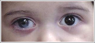
Phenotypic variation in ophthalmic manifestations of MIDAS syndrome (microphthalmia, dermal aplasia, and sclerocornea).
PETERS ANOMALY SKIN
Corneal pathology in microphthalmia with linear skin defects syndrome.

Although we do not have confirmation by histopathological examination of the patient’s corneas, the constellation of clinical findings most likely represent a variant of the microphthalmia with linear skin defects (MLS) syndrome.Ĭorneal opacification with multiple linear skin defects has been described as part of the microphthalmia with linear skin defects (MLS) syndrome, which is also known as the microphthalmia, dermal aplasia, sclerocornea (MIDAS) syndrome. 3 The dermatologic findings of congenital syphilis are most commonly a desquamative vesicular or maculopapular rash, 4 much different from the linear erythematous streaks seen in our patient. The corneal findings in congenital syphilis most often manifest as a nonulcerative interstitial keratitis with deep stromal blood vessel invasion, which usually presents later in life (around the age of 2 years or older). The dermatologic skin findings in our patient, along with the clinical manifestation of her corneas were not characteristic of congenital syphilis. 2 The patient was referred for further evaluation but unfortunately was lost to follow-up at our institution. 1 A B-scan ultrasound was done and her axial lengths were also decreased, measuring 13 mm in both eyes (normal is approximately 16-18 mm). On examination the patient was noted to have bilateral corneal opacification with mild clearing (Images A and B).Ĭ) External photograph revealing multiple linear reddish skin lesions overlying the faceĬorneal measurements performed at bedside with calipers measured decreased horizontal and vertical corneal diameters (8 and 7 millimeters (mm), respectively normal diameter is approximately 9.5-10.5 mm). She had an unremarkable birth history but her mother was noted to have positive syphilis serologies.
PETERS ANOMALY FULL
Secondary CORE Category: Pediatric Ophthalmology and Strabismus / Diseases of the Cornea, Anterior Segment and Irisĭiagnosis: Peter’s Anomaly with Microphthalmia and Linear Skin Defects SyndromeĪ 1 week old full term African-American female was evaluated for bilateral corneal clouding.

Keywords/Main Subjects: Cornea, corneal dystrophy, Peter’s anomaly, congenital corneal dystrophy Mifflin, MD 1ġ) University of Utah Moran Eye Center, Salt Lake City, UT Ģ) University of Texas Medical Branch, Galveston, TX Nakatsuka, MD 1,2 Mohamed Soliman, MD 2 Mark D. Title: Peter’s Anomaly with Microphthalmia and Linear Skin DefectsĪuthors: Austin S. Home / External Disease and Cornea / Basic and Clinical Concepts of Congenital Anomalies of the Cornea, Sclera, and Globe Peter’s Anomaly with Microphthalmia and Linear Skin Defects


 0 kommentar(er)
